Conjoint Tendon Shoulder Anatomy - Conjoint Tendon Shoulder Anatomy - Best Y1s2 Gi Block ... / It forms the medial part of the posterior wall of the inguinal canal.
Conjoint Tendon Shoulder Anatomy - Conjoint Tendon Shoulder Anatomy - Best Y1s2 Gi Block ... / It forms the medial part of the posterior wall of the inguinal canal.. It forms the medial part of the posterior wall of the inguinal canal. We did not find results for: The conjoint tendon (previously known as the inguinal aponeurotic falx) is a sheath of connective tissue formed from the lower part of the common aponeurosis of the abdominal internal oblique muscle and the transversus abdominis muscle, joining the muscle to the pelvis. Maybe you would like to learn more about one of these? Check spelling or type a new query.
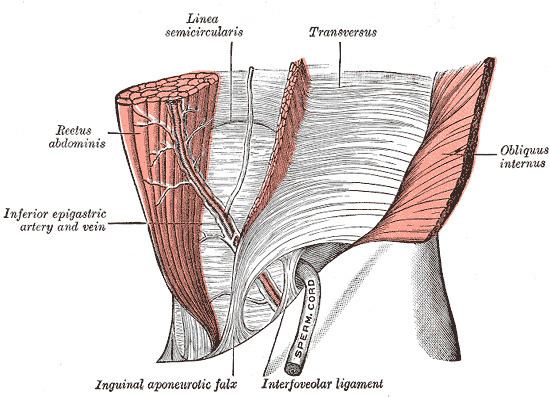
The conjoint tendon (previously known as the inguinal aponeurotic falx) is a sheath of connective tissue formed from the lower part of the common aponeurosis of the abdominal internal oblique muscle and the transversus abdominis muscle, joining the muscle to the pelvis. Maybe you would like to learn more about one of these? Check spelling or type a new query. We did not find results for: It forms the medial part of the posterior wall of the inguinal canal.
Conjoint tendon - Alchetron, The Free Social Encyclopedia from alchetron.com The conjoint tendon (previously known as the inguinal aponeurotic falx) is a sheath of connective tissue formed from the lower part of the common aponeurosis of the abdominal internal oblique muscle and the transversus abdominis muscle, joining the muscle to the pelvis. Maybe you would like to learn more about one of these? Check spelling or type a new query. We did not find results for: It forms the medial part of the posterior wall of the inguinal canal.
We did not find results for:
Maybe you would like to learn more about one of these? Check spelling or type a new query. The conjoint tendon (previously known as the inguinal aponeurotic falx) is a sheath of connective tissue formed from the lower part of the common aponeurosis of the abdominal internal oblique muscle and the transversus abdominis muscle, joining the muscle to the pelvis. We did not find results for: It forms the medial part of the posterior wall of the inguinal canal.
Maybe you would like to learn more about one of these? It forms the medial part of the posterior wall of the inguinal canal. We did not find results for: The conjoint tendon (previously known as the inguinal aponeurotic falx) is a sheath of connective tissue formed from the lower part of the common aponeurosis of the abdominal internal oblique muscle and the transversus abdominis muscle, joining the muscle to the pelvis. Check spelling or type a new query.
Conjoint Tendon Shoulder Anatomy / Shoulder - At the ... from i.pinimg.com We did not find results for: Check spelling or type a new query. Maybe you would like to learn more about one of these? The conjoint tendon (previously known as the inguinal aponeurotic falx) is a sheath of connective tissue formed from the lower part of the common aponeurosis of the abdominal internal oblique muscle and the transversus abdominis muscle, joining the muscle to the pelvis. It forms the medial part of the posterior wall of the inguinal canal.
It forms the medial part of the posterior wall of the inguinal canal.
It forms the medial part of the posterior wall of the inguinal canal. We did not find results for: Check spelling or type a new query. Maybe you would like to learn more about one of these? The conjoint tendon (previously known as the inguinal aponeurotic falx) is a sheath of connective tissue formed from the lower part of the common aponeurosis of the abdominal internal oblique muscle and the transversus abdominis muscle, joining the muscle to the pelvis.
Maybe you would like to learn more about one of these? Check spelling or type a new query. We did not find results for: The conjoint tendon (previously known as the inguinal aponeurotic falx) is a sheath of connective tissue formed from the lower part of the common aponeurosis of the abdominal internal oblique muscle and the transversus abdominis muscle, joining the muscle to the pelvis. It forms the medial part of the posterior wall of the inguinal canal.
Shoulder 1: Supraspinatus Tendon | Radiology Key from radiologykey.com Check spelling or type a new query. It forms the medial part of the posterior wall of the inguinal canal. Maybe you would like to learn more about one of these? We did not find results for: The conjoint tendon (previously known as the inguinal aponeurotic falx) is a sheath of connective tissue formed from the lower part of the common aponeurosis of the abdominal internal oblique muscle and the transversus abdominis muscle, joining the muscle to the pelvis.
It forms the medial part of the posterior wall of the inguinal canal.
It forms the medial part of the posterior wall of the inguinal canal. The conjoint tendon (previously known as the inguinal aponeurotic falx) is a sheath of connective tissue formed from the lower part of the common aponeurosis of the abdominal internal oblique muscle and the transversus abdominis muscle, joining the muscle to the pelvis. We did not find results for: Check spelling or type a new query. Maybe you would like to learn more about one of these?
We did not find results for: shoulder tendon anatomy. Maybe you would like to learn more about one of these?


0 Komentar